Diabetic Foot Ulcer Stages: Signs a Wound Is Getting Worse
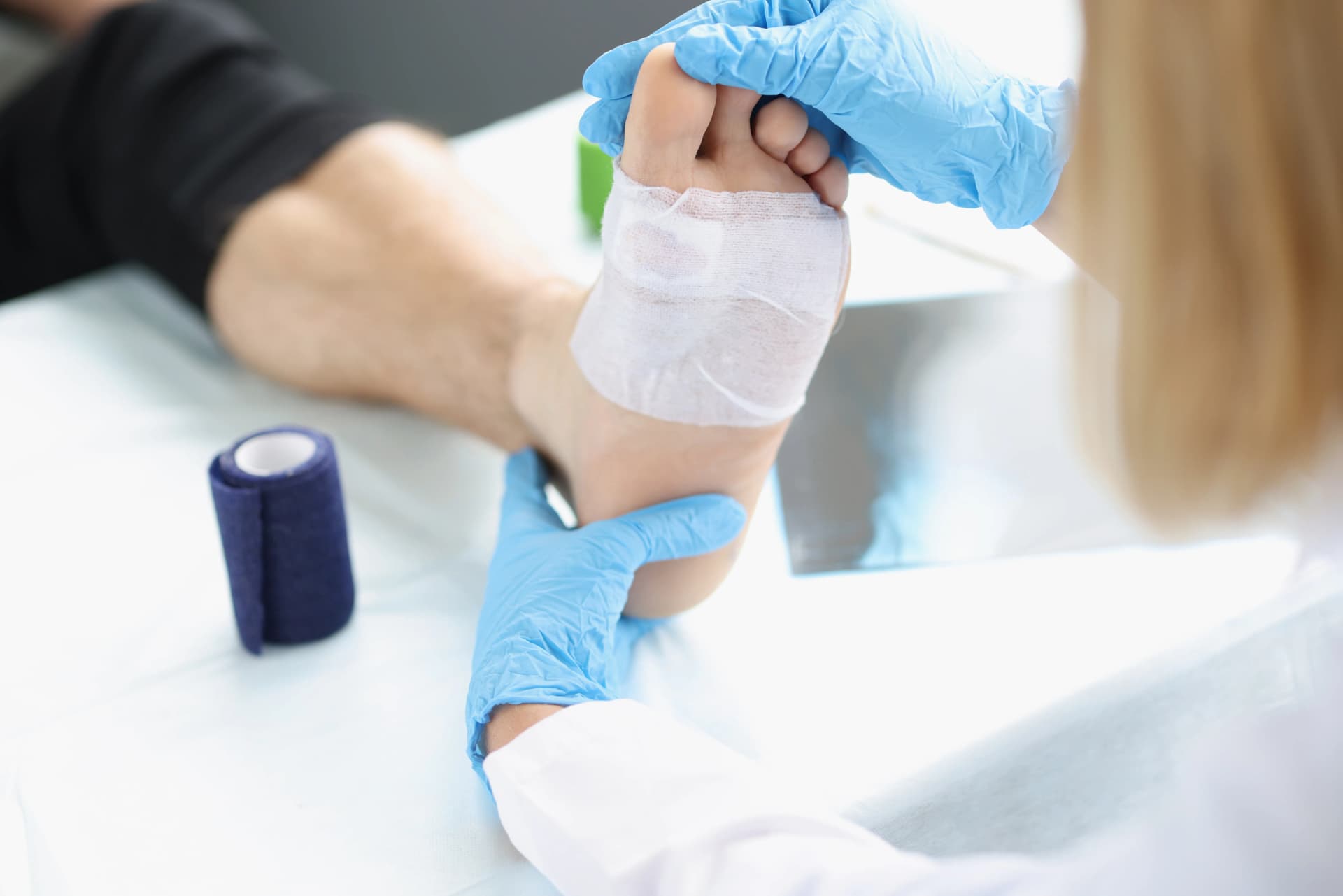
A diabetic foot wound can change quickly, sometimes without causing significant pain. Understanding the diabetic foot ulcer stages can help patients recognize concerning changes and seek timely medical care.
However, appearance alone cannot establish a wound’s severity. Healthcare professionals consider its depth, circulation, infection, tissue damage, and other clinical findings. Any new or worsening foot wound in someone with diabetes deserves professional evaluation.
What Is a Diabetic Foot Ulcer?
A diabetic foot ulcer is an open sore or wound that typically develops on the bottom, side, heel, or toes of the foot. It often begins when pressure, friction, a blister, or a minor injury damages the skin.
Diabetes can make these wounds more difficult to detect and treat. Diabetic neuropathy may reduce sensation, preventing someone from noticing pain or an injury. Poor circulation also limits the oxygen and nutrients required for tissue repair. Elevated blood glucose can impair immune function, increases the risk of infection, and prolongs healing time.
Patients with neuropathy, foot deformities, previous ulcers, kidney disease, or peripheral artery disease (PAD) are considered at particularly high risk. Even a small wound should be examined rather than treated as an ordinary cut or blister.
What Are the Stages of a Diabetic Foot Ulcer?
Clinicians use classification systems to describe diabetic foot wounds and guide the treatment plan. These systems consider factors such as wound depth, tissue involvement, infection, and reduced blood flow.
The Wagner system is commonly discussed with patients. It uses grades from 0 through 5:
Grade 0: No open ulcer, but the foot may be at risk.
Grade 1: A superficial ulcer involving the skin.
Grade 2: A deeper ulcer reaching a tendon, joint capsule, or other underlying structure.
Grade 3: A deep wound with an abscess, bone infection, or joint infection.
Grade 4: Localized gangrene affecting part of the foot.
Grade 5: Extensive gangrene involving most or all of the foot.
These grades should not be interpreted as a timeline every wound will follow. A clinician must classify the ulcer because its appearance may not reveal infection, circulation problems, or deeper tissue involvement. Current clinical guidance also recommends classification systems that account for site, area, depth, infection and sensation.
Early-Stage Diabetic Foot Ulcers
An early ulcer may appear as a shallow break in the skin. Patients might notice a blister, drainage on a sock, a callus with discoloration underneath, or a small crater-like area.
Although the wound may look minor, pressure can continue damaging the tissue each time the person stands or walks. Reduced sensation may also make the ulcer painless. Early medical care can address pressure, wound protection, circulation, glucose management, and infection risk before deeper structures become affected.
Patients should not cut away calluses, drain blisters, or apply harsh antiseptics without clinical guidance.
Deeper or More Advanced Wounds
As an ulcer progresses, it can extend through the skin into fatty tissue, tendons, muscles, joints, or bone. The wound may become wider or deeper, produce more drainage, develop dark tissue, or expose underlying structures.
Advanced ulcers often require coordinated wound care. Treatment may include pressure relief, debridement, dressings, infection management, circulation testing, glucose control, and nutritional support. Reduced blood supply caused by peripheral artery disease (PAD) may require vascular assessment or intervention.
Severely damaged or infected tissue can require surgical intervention. Prompt treatment supports healing wounds while helping prevent infection, hospitalization, or amputation.
When Infection Becomes a Concern
Healthcare professionals diagnose diabetic foot infection through clinical findings rather than wound appearance alone. Possible signs of infection include:
New or spreading redness
Swelling or warmth around the wound
Increasing or foul-smelling drainage
Pus
New tenderness or pain
Tissue discoloration or breakdown
Fever, chills, weakness, or confusion
Neuropathy and impaired immune responses can make some symptoms less noticeable. A patient may not experience intense pain or fever even when an infection is serious. Spreading redness, black tissue, systemic illness, or rapidly worsening swelling warrants urgent medical attention.
Signs a Diabetic Foot Ulcer May Be Getting Worse
A worsening wound does not always cause dramatic symptoms. Patients and caregivers should pay attention to changes between professional evaluations, including:
The ulcer becoming wider, deeper, or more difficult to see beneath a callus
Increased drainage or blood on socks, shoes, or dressings
New odor, pus, redness, swelling, or warmth
Skin around the wound becoming pale, blue, purple, or black
Exposed fat, tendon, joint, or bone
New numbness or changes in sensation
A foot becoming unusually cool
Red streaks extending from the wound
Fever, chills, confusion, weakness, or elevated blood glucose
These changes do not confirm a particular stage, but they may indicate impaired circulation, deeper damage, or infection. Contact a medical professional promptly rather than waiting for a scheduled visit. Severe discoloration, rapidly spreading redness, systemic symptoms, or suspected gangrene requires urgent evaluation.
Why Some Diabetic Foot Wounds Don’t Heal
Normal healing depends on blood flow, oxygen, immune activity, pressure control, and the body’s ability to rebuild damaged tissue. Diabetes can disrupt several of these processes simultaneously.
Common barriers include:
Neuropathy that allows repeated pressure or unnoticed injuries
Poorly controlled blood glucose
Continued walking or standing on the affected area
Poor circulation associated with PAD
Infection or dead tissue
Swelling
Inadequate wound care
Smoking
Poor nutrition
Kidney disease or other health conditions
Footwear that continues rubbing or compressing the wound
Effective care addresses more than the visible opening. A comprehensive treatment plan may involve offloading, regular wound measurement, debridement, infection treatment, circulation evaluation, glucose management, appropriate dressings, and specialist referrals.
No single therapy replaces these fundamentals. Coordinated care offers the best opportunity for healing faster, limiting deeper tissue damage, and achieving improved healing and reduced complication risks.
When Is HBOT Considered for a Diabetic Foot Ulcer?
Hyperbaric oxygen therapy may be considered as an adjunctive treatment for certain advanced diabetic foot ulcers that have not responded adequately to standard wound care. During HBOT, a patient breathes oxygen inside a pressurized chamber, increasing the amount of oxygen available to affected tissues.
HBOT does not replace offloading, glucose management, debridement, infection treatment, vascular care, or necessary surgery. Eligibility depends on the wound’s severity, blood supply, medical history, previous treatment, and whether the patient can safely undergo therapy.
Medicare coverage criteria, for example, generally describe HBOT as an adjunct for Wagner grade 3 or higher lower-extremity diabetic wounds that show no measurable healing after at least 30 days of standard wound therapy. Individual clinical recommendations and insurance requirements may differ.
Patients can learn more about Hyperbaric Oxygen Therapy for Diabetic Wound Healing and discuss whether it may support their broader care plan.
Frequently Asked Questions
Can a diabetic foot ulcer heal?
Yes. Many diabetic foot ulcers can heal with early, coordinated care. Wound depth, circulation, infection, glucose management, pressure relief, nutrition, and other health conditions influence the likely healing time and treatment required.
How do you know if a diabetic foot ulcer is infected?
Redness, warmth, swelling, pus, odor, increased drainage, pain, or rapidly deteriorating tissue may be concerning. Because symptoms can be subtle, a healthcare professional should evaluate suspected infection rather than relying on self-diagnosis.
When should you see a doctor for a diabetic foot wound?
Contact a medical professional when any blister, sore, crack, discoloration, or open wound develops. Seek prompt care for worsening drainage, swelling, redness, dark tissue, fever, chills, weakness, or other concerning changes.
Does every diabetic foot ulcer need HBOT?
No. HBOT is generally reserved for appropriately selected wounds and is used alongside comprehensive wound care. A qualified clinician must evaluate the ulcer, circulation, treatment history, overall health, and possible risks before recommending it.
Explore Your Options for a Non-Healing Diabetic Wound
If a diabetic foot wound is not improving or you have questions about HBOT, contact NorCal Hyperbarics to request an evaluation and learn whether adjunctive hyperbaric care may be appropriate for your treatment plan.
Call us: (925) 555-0180 | Concord, CA | www.norcalhbo.com
Advanced Healing & Wellness
How To Get Started with Your Tailored Hyperbaric Oxygen Therapy Sessions
From comprehensive assessments to personalized treatments and ongoing support, our approach ensures that every stage is aligned with your specific needs, helping you heal faster, feel stronger, and achieve lasting results.
Thorough Health Assessments to Understand Your Unique Needs

Developing a Hyperbaric Oxygen Therapy Regimen Just for You

Providing Support Throughout Your Healing Journey

Comprehensive Solutions for Lasting Health and Wellness
Discover our wide range of targeted therapies designed to enhance well-being, address chronic conditions, and support optimal health. Each treatment is customized to meet your unique needs, promoting recovery and vitality.


Begin Your Healing Journey with NorCal Hyperbarics
Declining health isn’t permanent. Whether addressing specific challenges or age-related changes, NorCal Hyperbaric’s personalized therapy can enhance your quality of life.

